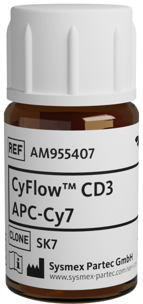
CyFlow™ CD3 APC-Cy7
| Alternative Name: | Leu4, T3 |
| Antigen: | CD3 |
| Application: | Flow cytometry |
| Clonality: | monoclonal |
| Clone: | SK7 |
| Emission Maximum: | 780 nm |
| Excitation Maximum: | 650 nm, 750 nm |
| Field of Interest: | Immunophenotyping |
| Format/Fluorochrome: | APC-Cy7 |
| Isotype: | IgG1 |
| Laser: | Red |
| Regulatory Status: | CE IVD |
| Source Species: | Mouse |
| Target Species: | Human |
| Product number: | AM955407 |
CE IVD
| HLDA Workshop | HLDA II—WS Code T118 HLDA III—WS Code T492 |
| Concentration Unit | µg/mL |
| Concentration | 25 |
| Quantity | 100 tests |
| Volume | 1.0 mL |
| Immunogen | Human thymocytes |
| Background Information | CD3 complex is crucial in transducing antigen-recognition signals into the cytoplasm of T cells and in regulating the cell surface expression of the TCR complex. T cell activation through the antigen receptor (TCR) involves the cytoplasmic tails of the CD3 subunits CD3γ, CD3 δ, CD3ε and CD3ζ. These CD3 subunits are structurally related members of the immunoglobulins superfamily encoded by closely linked genes on human chromosome 11. The CD3 components have long cytoplasmic tails that associate with cytoplasmic signal transduction molecules. This association is mediated at least in part by a double tyrosine-based motif present in a single copy in the CD3 subunits. CD3 may play a role in TCR-induced growth arrest, cell survival and proliferation. The CD3 antigen is present on 68-82% of normal peripheral blood lymphocytes, 65-85% of thymocytes and Purkinje cells in the cerebellum. It is never expressed on B or NK cells. Decreased percentages of T lymphocytes may be observed in some autoimmune diseases. |
| Antigen Distribution | CD3 (T3, Leu4) is expressed on peripheral blood lymphocytes, thymocytes and Purkinje cells in the cerebellum; it is lost on B or NK cells. CD3 complex is crucial in transducing antigen-recognition signals into the cytoplasm of T cells and in regulating the cell surface expression of the TCR complex. T cell activation through the antigen receptor (TCR) involves the cytoplasmic tails of the CD3 subunits CD3γ, CD3δ, CD3ε and CD3ζ. |
| Usage | CE IVD usage Placeholder |
| Storage Buffer | The reagent is provided in stabilizing phosphate buffered saline (PBS) solution, pH ≈7.4, containing 0.09% (w/v) sodium azide and 0.2% (w/v) BSA. |
| Storage | Avoid prolonged exposure to light. Store in the dark at 2-8°C. Do not freeze. |
| Stability | Do not use after expiration date stamped on vial label. |
| Wood GS, Burns BF, Dorfman RF, Warnke RA: The immunohistology of non‑T cells in the acquired immunodeficiency syndrome. Am J Pathol. 1985 Sep; 120(3):371‑9. < PMID: 3898858 > | Li B, Wang H, Dai J, Ji J, Qian W, Zhang D, Hou S, Guo Y: Construction and characterization of a humanized anti‑human CD3 monoclonal antibody 12F6 with effective immunoregulation functions. Immunology. 2005 Dec; 116(4):487‑98. < PMID: 16313362 > | Recio MJ, Moreno-Pelayo MA, Kiliç SS, Guardo AC, Sanal O, Allende LM, Pérez-Flores V, Mencía A, Modamio-Høybjør S, Seoane E, Regueiro JR: Differential biological role of CD3 chains revealed by human immunodeficiencies. J Immunol. 2007 Feb 15; 178(4):2556‑64. < PMID: 17277165 > | Guttman-Yassky E, Vugmeyster Y, Lowes MA, Chamian F, Kikuchi T, Kagen M, Gilleaudeau P, Lee E, Hunte B, Howell K, Dummer W, Bodary SC, Krueger JG: Blockade of CD11a by efalizumab in psoriasis patients induces a unique state of T‑cell hyporesponsiveness. J Invest Dermatol. 2008 May; 128(5):1182‑91. < PMID: 18239614 >