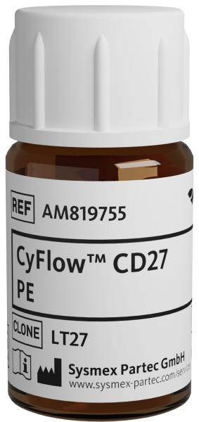
CyFlow™ CD27 PE

| Alternative Name: | S152, T14 , TNFRSF7, TP55 |
| Antigen: | CD27 |
| Application: | Flow cytometry |
| Clonality: | monoclonal |
| Clone: | LT27 |
| Emission Maximum: | 576 nm |
| Excitation Maximum: | 496 nm, 565 nm |
| Field of Interest: | Immunophenotyping |
| Format/Fluorochrome: | PE |
| Isotype: | IgG2a |
| Laser: | Blue , Green, Yellow |
| Regulatory Status: | CE IVD |
| Source Species: | Mouse |
| Target Species: | Human |
| Product number: | AM819755 |
CE IVD
| HLDA Workshop | HLDA V—WS Code T T-CD27.01 |
| Concentration Unit | µg/mL |
| Concentration | 10 |
| Quantity | 100 tests |
| Volume | 1.0 mL |
| Immunogen | Human peripheral blood lymphocytes |
| Background Information | CD27 is a transmembrane 55 kDa protein of the nerve growth factor-receptor family, expressed as a disulfide-linked homodimer on mature thymocytes, peripheral blood T cells and a subpopulation of B cells. Activation of T cells via TCR-CD3 complex results in upregulation of CD27 expression on the plasma membrane as well as in the release of its soluble 28-32 kDa form, sCD27, detected in the plasma, urine or spinal fluid. This sCD27 is an important prognostic marker of acute and chronic B cell malignancies. RgpA, a cystein proteinase, although activating T cells through the protease-activated receptors (PARs), degradates CD27 and counteracts T cell activation mediated by CD27 and its ligand CD70. |
| Antigen Distribution | CD27 (T14, S152, TP55, TNFRSF7) is a 55 kDa type I transmembrane glycoprotein (member of the TNF-receptor superfamily) expressed as a disulfide-linked homodimer on mature thymocytes, peripheral blood T cells and a subpopulation of B cells and NK cells. Activation of T cells via TCR-CD3 complex results in upregulation of CD27 expression on the plasma membrane as well as in the release of its soluble 28-32 kDa form, sCD27, detected in the plasma, urine or spinal fluid. |
| Usage | CE IVD usage Placeholder |
| Storage Buffer | The reagent is provided in stabilizing phosphate buffered saline (PBS) solution, pH ≈7.4, containing 0.09% (w/v) sodium azide and 0.2% (w/v) BSA. |
| Storage | Avoid prolonged exposure to light. Store in the dark at 2-8°C. Do not freeze. |
| Stability | Do not use after expiration date stamped on vial label. |
| Doussis IA, Gatter KC, Mason DY: CD68 reactivity of non‑macrophage derived tumours in cytological specimens. J Clin Pathol. 1993 Apr; 46(4):334‑6. < PMID: 7684403 > | van Oers MH, Pals ST, Evers LM, van der Schoot CE, Koopman G, Bonfrer JM, Hintzen RQ, von dem Borne AE, van Lier RA: Expression and release of CD27 in human B‑cell malignancies. Blood. 1993 Dec 1; 82(11):3430‑6. < PMID: 8241510 > | Molica S, Vitelli G, Levato D, Crispino G, Dell'Olio M, Dattilo A, Matera R, Gandolfo GM, Musto P: CD27 in B‑cell chronic lymphocytic leukemia: Cellular expression, serum release and correlation with other soluble molecules belonging to nerve growth factor receptors (NGFr) superfamily. Haematologica. 1998 May; 83(5):398‑402l. < PMID: 9658722 > | Kara IO, Sahin B, Gunesacar R: Expression of soluble CD27 and interleukins‑8 and ‑10 in B‑cell chronic lymphocytic leukemia: correlation with disease stage and prognosis. Adv Ther. 2007 Jan-Feb; 24(1):29‑40. < PMID: 17526459 > | Yun LW, Decarlo AA, Hunter N: Blockade of protease‑activated receptors on T cells correlates with alterd proteolysis of CD27 by gingipains of Porphyromonas gingivalis. Clin Exp Immunol. 2007 Nov; 150(2):217‑29. < PMID: 17937677 > | Kanderova V, Kuzilkova D, Stuchly J, Vaskova M, Brdicka T, Fiser K, Hrusak O, Lund‐Johansen F, Kalina T: High‐resolution antibody array analysis of childhood acute leukemia cells. Mol Cell Proteomics. 2016 Apr 1; 15(4):1246‐61. < PMID: 26785729 >
